| Theories and Techniques of Oral Implantology (vol.1) (published 1970) | Dr. Leonard I. Linkow |
|
|
Next Page |
| This is an archival HTML version of this book originally hosted here in 2006. The HTML may not display well on modern browsers. Please view the modern PDF Version for a better viewing experience. |
Implant histology 127
Center, San Juan, Puerto Rico, extensively studied the specimen, both macroscopically and microscopically.
Bodine and Mohammed found that the metal structure had remained flush with the bone and that there was no overhang below the mylohyoid ridge. Bone completely covered the implant's lingual portion in the right posterior quadrant in two areas: the mandibular torus and just distal to the anterior abutment from a point midway between the abutments and extending to the posterior abutment. Over almost the entire mandible was a thin, whitish, semi-transparent soft tissue.
For more extensive study the mandible with the implant in place was divided near the midline, and these halves were again divided midway between the two abutment posts. Of the resulting four segments, that containing the right posterior abutment was selected for serial cross sectioning. Bodine and Mohammed removed the implant and decalcified the bone in a 5% nitric acid solution over a period of several months.
A longitudinal section made through the most posterior extent of the peripheral frame, well away from the abutment post, shows clearly where the em-bedded metal was (Fig. 4-97). Collagenous fibrous connective tissue, about 0.2 to 0.5 mm. thick, lies between the metal space and the healthy normal
bone. There is no epithelial tissue. The collagenous tissue is healthy with no evidence of inflammation, although the first few layers of its cells are in contact with metal and are compressed. These cells, upon higher magnification (Fig. 4-98) were found to resemble those usually found only in tendons and ligaments.
In another section, made buccolingually midway bet een the abutment and the most posterior portion of the implant (Fig. 4-99), bone extends slightly up-ward between the metal struts. There is no evidence of inflammation or epithelial imagination along the spaces occupied by the implant. The connective tissue is healthy and once again exhibits the compactness of those cells closest to the metal. This tendon-like characteristic was found in all slides, and Bodine and Mohammed concluded that this was typical around the implant.
Midway between the right posterior and anterior abutments another section was made (Fig. 4-100). There was no evidence of epithelial cells or inflammation. Between metal and bone appeared a 0.25-mm. band of connective tissue; again the cell layers closest to the metal were tendon-like.
The bone observed macroscopically to have grown in two areas over the lingual section of the implant was closely examined (Fig. 4-101) . It contained osteoblasts, lamellae, and haversian systems characteristic
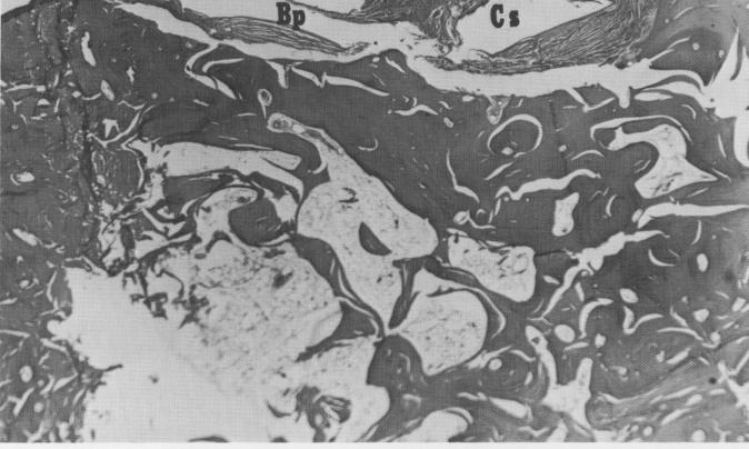
Fig. 4-99. In a section made midway between the abutment post and the most posterior part of the implant the sites of two struts are demarcated by firm, healthy connective tissue. Also, a small hill of bone appears between the central secondary strut site (Cs) and the buccal peripheral frame site (Bp). (From Bodine, R. L., Jr., and Mohammed, C. I.: Histologic studies of a human mandible supporting an implant denture, J. Prosth. Dent. 21 [2] :203-216, 1969.)
|
|
Page 127 |
Next Page |
|
Copyright warning: This information is presented here for free for anyone to study online. We own exclusive internet copyrights on all content presented on this website. We use sophisticated technology to identify and legally close down websites that reproduce copyrighted content without permission - so please don’t do it.
|