| Theories and Techniques of Oral Implantology (vol.2) (published 1970) | Dr. Leonard I. Linkow |
|
|
Next Page |
| This is an archival HTML version of this book originally hosted here in 2006. The HTML may not display well on modern browsers. Please view the modern PDF Version for a better viewing experience. |
618 Theories and techniques of oral implantology
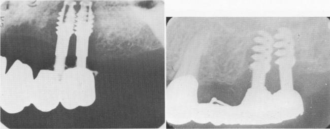
Fig. 14-19. These two radiographs reveal a rapid resorption of bone because of the proximity of the implants. They should be set at least 4 mm. away from one another.
A
C
B
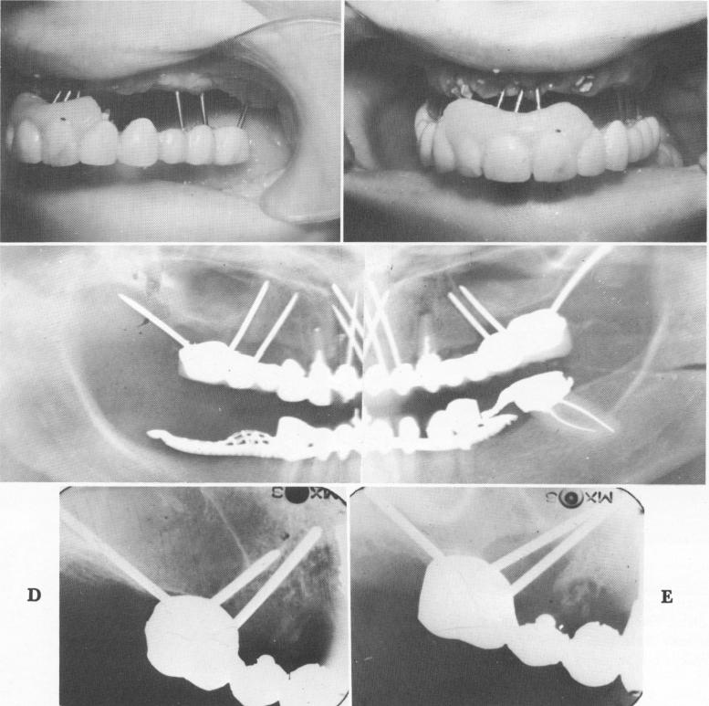
Fig. 14-20. A and B, Often a bridge can be removed with all of the triplant pins still attached to it and still diverging from each other. Very seldom will any fibrous tissue be attached to the pins, such as is usually the case with the post type and blade implants, indicating the lack of a physiologic pseudomembrane attachment. C, A Panorex showing the entire prosthesis and the diverging pins just prior to its removal. D, Periapical radiograph taken immediately after pins were inserted. E, Six months' postoperative radiograph reveals large breakdown of bone around the two mesially directed pins, which were placed too close together and too close to the buccal plate of bone.
|
|
Page 618 |
Next Page |
|
Copyright warning: This information is presented here for free for anyone to study online. We own exclusive internet copyrights on all content presented on this website. We use sophisticated technology to identify and legally close down websites that reproduce copyrighted content without permission - so please don’t do it.
|