| Theories and Techniques of Oral Implantology (vol.1) (published 1970) | Dr. Leonard I. Linkow |
|
|
Next Page |
| This is an archival HTML version of this book originally hosted here in 2006. The HTML may not display well on modern browsers. Please view the modern PDF Version for a better viewing experience. |
128 Theories and techniques of oral implantology
Fig. 4-100. The material midway between the right posterior and anterior abutments was sectioned. Two sites are shown surrounded by dense connective tissue, the lingual peripheral frame site (L) and the buccal secondary strut site (Bs). The crest of the ridge is marked R and the bone over the lingual frame site, B. (From Bodine, R. L., Jr., and Mohammed, C. I.: Histologic studies of a human mandible supporting an implant denture, J. Prosth. Dent. 21 [2] : 203-216, 1969.)
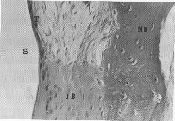
Fig. 4-101. Magnification of box in Fig. 4-100. Upon magnification of the bone over the lingual frame site (S), its character becomes clearer. There is mature bone (MB) separated from the implant site by typical connective tissue (T), and immature bone contacting the implant site (IB). (From Bodine, R. L., Jr., and Mohammed, C. I.: Histologic studies of a human mandible supporting an implant denture, J. Prosth. Dent. 21: [2] :203-216, 1969.)

|
|
Page 128 |
Next Page |
|
Copyright warning: This information is presented here for free for anyone to study online. We own exclusive internet copyrights on all content presented on this website. We use sophisticated technology to identify and legally close down websites that reproduce copyrighted content without permission - so please don’t do it.
|