| Theories and Techniques of Oral Implantology (vol.2) (published 1970) | Dr. Leonard I. Linkow |
|
|
Next Page |
| This is an archival HTML version of this book originally hosted here in 2006. The HTML may not display well on modern browsers. Please view the modern PDF Version for a better viewing experience. |
620 Theories and techniques of oral implantology
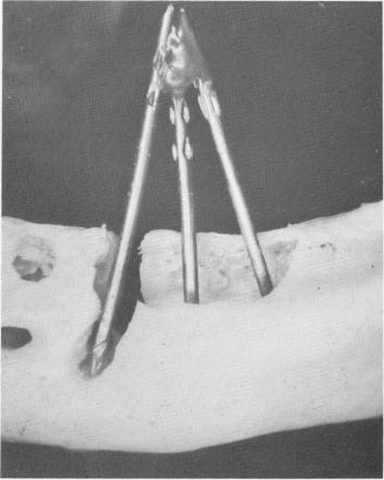
Fig. 14-24. A, A good portion of the alveolar bone was re-moved from a mandible and a triplant was placed into the open cavity. B, A lateral view showing the pins in the void and the labial and lingual cortical plates of bone still intact.
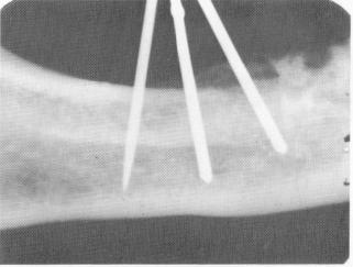
Fig. 14-25. The dried specimen was radiographed and the x-ray shows the pins to be buried in bone. The true situation was camouflaged by the buccal and lingual plates of cortical bone.
Fig. 14-26. The buccal and lingual cortical plates were then perforated a few millimeters in width and 1/2 inch in length, and once again the pins were placed inside.
are characterized by their smooth surfaces. This means that, unlike the post type implants and the blade-vent, no pseudoperiodontal membrane is found to encourage osteogenesis. Collagenous tissue does grow up to and encircle the pins, but it forms a kind of sleeve through which the pins can slip without pulling on the bone. As the bone around the pins is not stimulated, a certain amount of resorption occurs. This tendency becomes severe if the triplant is not securely stabilized.
Formerly, when patients complained of pain or mobility, the implantologist could find little reason for it on x-ray studies. The following studies show that diagnosis solely by x-ray can be misleading. Linkow demonstrated experimentally with mandibular and maxillary jaws that the density of the buccal, lingual, or palatal cortical plates of bone can disguise the true resorption picture taking place in alveolar bone flanking a narrow pin. Scooping out alveolar bone, he placed the pins inside the hole (Fig. 14-24). Then he x-rayed the dried specimen (Fig. 14-25). Because the cortical plates of bone camouflaged the lack of alveolar bone, bone appears to completely encapsulate the triplant. He then cut away portions of cortical plates (Fig. 14-26) and once again radiographed the area (Fig. 14-27). In this way he established the fallibility of x-rays in diagnosing bone resorption in triplants.
It is now known that each triplant used in long

|
|
Page 620 |
Next Page |
|
Copyright warning: This information is presented here for free for anyone to study online. We own exclusive internet copyrights on all content presented on this website. We use sophisticated technology to identify and legally close down websites that reproduce copyrighted content without permission - so please don’t do it.
|