| Theories and Techniques of Oral Implantology (vol.1) (published 1970) | Dr. Leonard I. Linkow |
|
|
Next Page |
| This is an archival HTML version of this book originally hosted here in 2006. The HTML may not display well on modern browsers. Please view the modern PDF Version for a better viewing experience. |
124 Theories and techniques of oral implantology
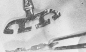
Bone had completely encapsulated the implant all along the buccal surface of the blade (Fig. 4-90). It would have also appeared along its lingual facing if it hadn't been chipped away with the chisel and mallet.
When the lingual surface was studied, bone growth through the open vents was clearly acknowledged (Fig. 4-91).
Fig. 4-90. The implant was removed with a massive amount of bone.
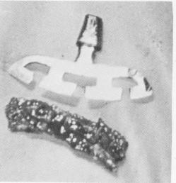
After the hone block was separated from the blade implant, bone regeneration to the exact con-figuration of the open vents was without doubt recognizable (Fig. 4-92). When the bone was viewed from a lateral view looking down upon it, it was clearly evident where the bone had grown through the open areas of the blade (Fig. 4-93) .
Another blade was immediately replaced 3 mm. lingual to where it was just removed (Fig. 4-94).
SUBPERIOSTEAL IMPLANTS
Because subperiosteal implants sit on, rather than in, bone, the majority of histologic studies on them concern the nature of the soft tissues lying over the implant and around its abutment posts. However, studies on possible changes in the bone caused by trauma or the presence of a foreign material have also been undertaken.
Herschfus on experimental implantations
Leon Herschfus, D.D.S., reported his experiments with subperiosteal implants in mongrel dogs in the
Fig. 4-91. Viewing the blade from its lingual surface where the bone was cut away, it was clearly evident that bone had grown through the open vents.
Fig. 4-93. Observation of the bone from a lateral view clearly illustrates the unrestricted growth of bone through the blade-vents.
Fig. 4-92. The surface of the bone in direct contact with the buccal surface of the blade shows exact details of bone growth following the outline of the openings in the blade.
Fig. 4-94. The implant was reinserted lingual to the original implant site.


|
|
Page 124 |
Next Page |
|
Copyright warning: This information is presented here for free for anyone to study online. We own exclusive internet copyrights on all content presented on this website. We use sophisticated technology to identify and legally close down websites that reproduce copyrighted content without permission - so please don’t do it.
|