| Theories and Techniques of Oral Implantology (vol.1) (published 1970) | Dr. Leonard I. Linkow |
|
|
Next Page |
| This is an archival HTML version of this book originally hosted here in 2006. The HTML may not display well on modern browsers. Please view the modern PDF Version for a better viewing experience. |
Implant histology 123
the presence of epithelium. Upon discussion of the results with an associate pathologist, it was concluded that the inflammation was relatively insignificant. The "granular black foreign material" provoking the foreign body reaction was clearly not part of the implant but may have entered the site during the operator), procedures. As for the small epithelial fragment, it may have been lodged there while trying to remove the implant. The most significant finding, the pathologists agreed, was the fact that bone bridges did grow through the vents in the implant.
Cullen on the Linkow blade
In November. 1969, a blade implant was placed in the left edentulous posterior area of the mandible of a 50-year-old woman at the Royal Society of Medicine in London, England.
Six months later, in May, 1970, it was decided to remove the well-functioning implant only because it was originally placed too far buccally and had been
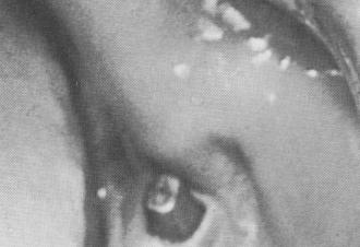
Fig. 4-86. The inside of the cheek followed the outline of the post.
Fig. 4-87. An incision was made in preparation for removal of the implant.
irritating the inside of the patient's cheek (Figs. 4-85 and 4-86).
An incision was made along the fibromucosal tissue above the underlying blade (Fig. 4-87). When the tissues were retracted it was noticed that the superior surface of the shoulders of the implant were still visible (Fig. 4-88).
Dr. Cullen tried unsuccessfully to remove the implant with a flat chisel by wedging the instrument between the hone flanking the blade on its lingual surface (Fig. 4-89). He then decided to bur the bone away from the mesial and distal proxiutal surfaces of the blade. It was then that he first realized that bone grew over the mesial and distal portions of the shoulder, which prevented dislodging the implant:. Not until this bone was completely eliminated could the implant be removed. It still took a great deal of careful surgery using the chisel as a wedge between the lingual bone and the lingual surface of the blade to remove the implant.
Fig. 4-88. The tissues were retracted to expose the under-lying bone.
Fig. 4-89. The removal of the implant was attempted by wedging a chisel between the implant and the bone on its lingual surface.


|
|
Page 123 |
Next Page |
|
Copyright warning: This information is presented here for free for anyone to study online. We own exclusive internet copyrights on all content presented on this website. We use sophisticated technology to identify and legally close down websites that reproduce copyrighted content without permission - so please don’t do it.
|